


Home / Training / Manuals / Atlas of breast cancer early detection / Cases
Atlas of breast cancer early detection
Go back to the list of case studies
.png) Click on the pictures to magnify and display the legends
Click on the pictures to magnify and display the legends
| Case number: | 054 |
| Age: | 62 |
| Clinical presentation: | Postmenopausal woman with average risk of developing breast cancer presented with right nipple retraction of duration 4 months. She had also noted a lump in the right breast a month before presentation. Examination revealed a hard lump, 6 cm in diameter, fixed to the rest of the breast tissue but not to the underlying muscles. Axilla was unremarkable. |
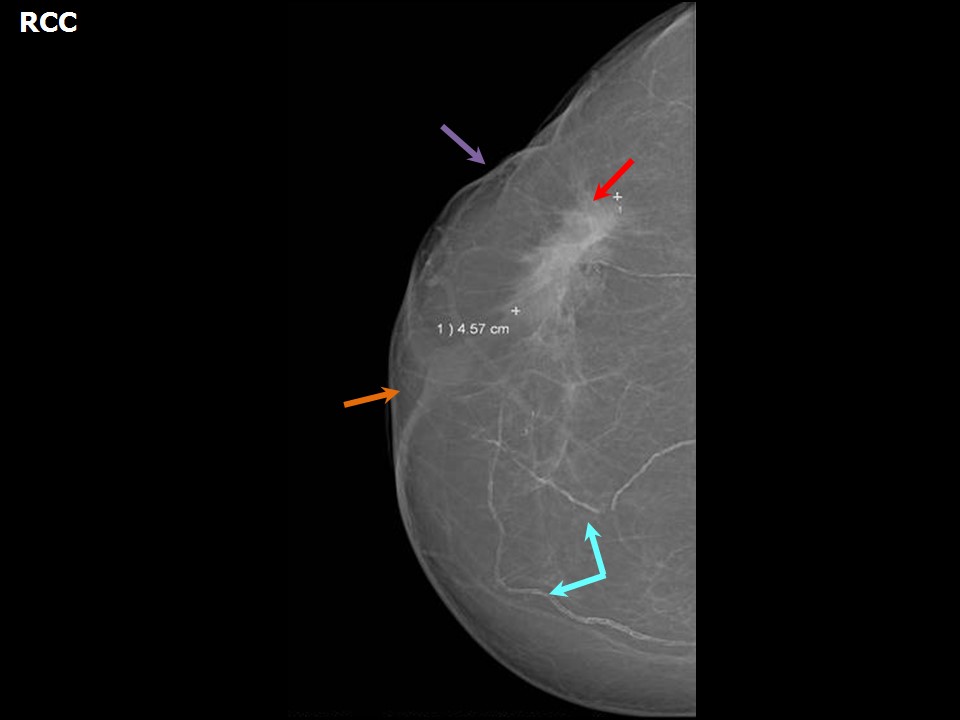
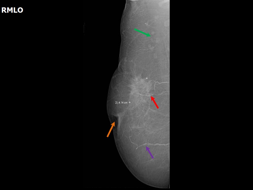
Mammography:
 |  |
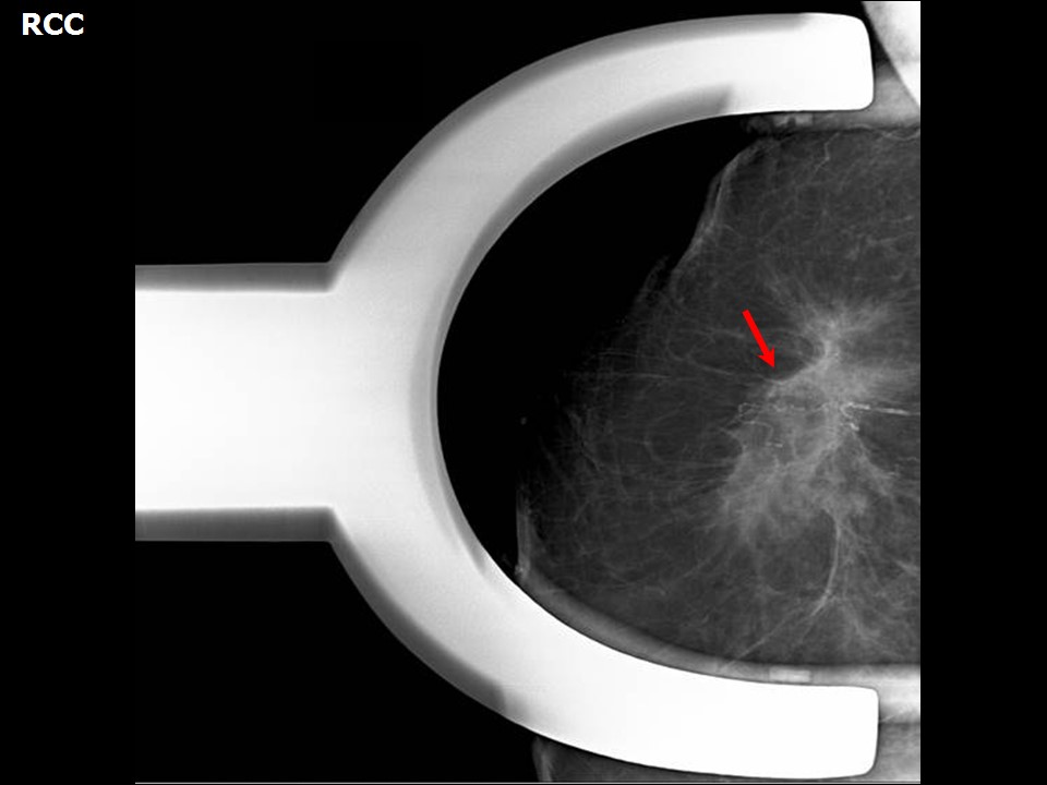 |
| Breast composition: | ACR category a (the breasts are almost entirely fatty) | Mammography features: |
| ‣ Location of the lesion: | Right breast, upper outer quadrant at 1011 oclock, middle third |
| ‣ Mass: | |
| • Number: | 1 |
| • Size: | 4.6 × 4.1 cm |
| • Shape: | Irregular |
| • Margins: | Spiculated |
| • Density: | High |
| ‣ Calcifications: | |
| • Typically benign: | Vascular calcification |
| • Suspicious: | None |
| • Distribution: | None |
| ‣ Architectural distortion: | Present |
| ‣ Asymmetry: | None |
| ‣ Intramammary node: | None |
| ‣ Skin lesion: | None |
| ‣ Solitary dilated duct: | None |
| ‣ Associated features: | Skin retraction and nipple retraction |
Ultrasound:
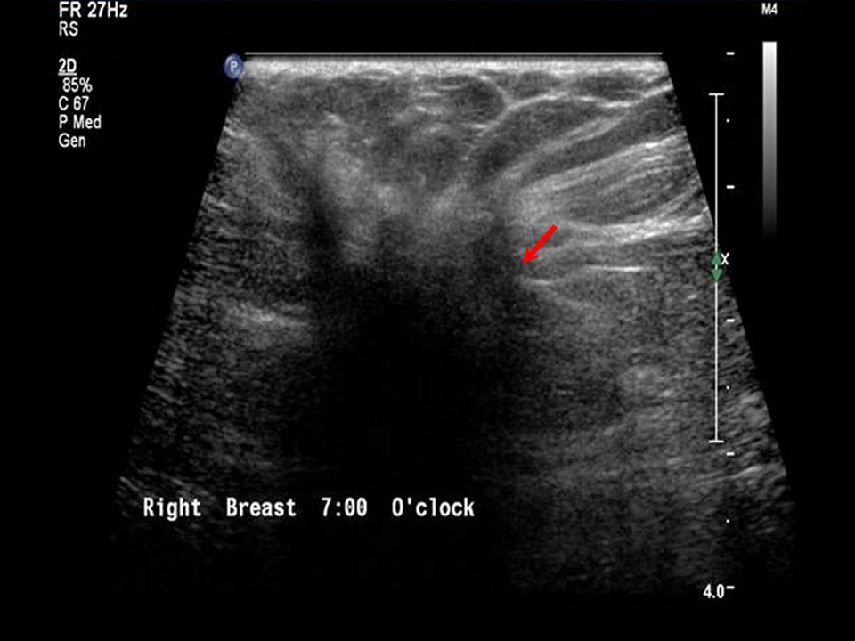 | 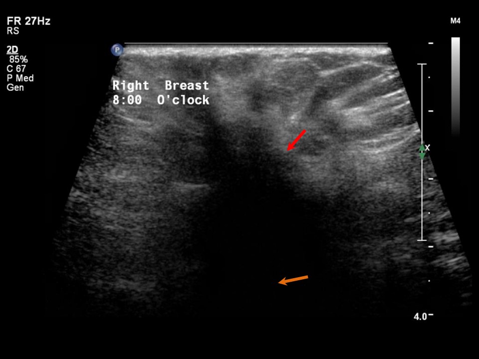 |
| Ultrasound features: Right breast, upper outer quadrant at 10 oclock position | |
| ‣ Mass | |
| • Location: | Right breast, upper outer quadrant at 10 oclock position |
| • Number: | 1 |
| • Size: | > 3.0 cm in greatest dimension |
| • Shape: | Irregular |
| • Orientation: | Not parallel |
| • Margins: | Spiculated |
| • Echo pattern: | Hypoechoic |
| • Posterior features: | Posterior shadowing |
| ‣ Calcifications: | None |
| ‣ Associated features: | Architectural distortion and skin retraction |
| ‣ Special cases: | None |
BI-RADS:
BI-RADS Category: 5 (highly suggestive of malignancy)Further assessment:
Further assessment advised: Referral for cytologyCytology:
 |
| Cytology features: | |
| ‣ Type of sample: | FNAC |
| ‣ Site of biopsy: | |
| • Laterality: | Right |
| • Quadrant: | Upper outer |
| • Localization technique: | Palpation |
| • Nature of aspirate: | whitish |
| ‣ Cytological description: | Smears show mainly dispersed malignant cells. These cells have a high N:C ratio. Many cells show small cytoplasmic vacuoles in the cytoplasm |
| ‣ Reporting category: | Malignant |
| ‣ Diagnosis: | Carcinoma |
| ‣ Comments: | None |
Histopathology:
MRM
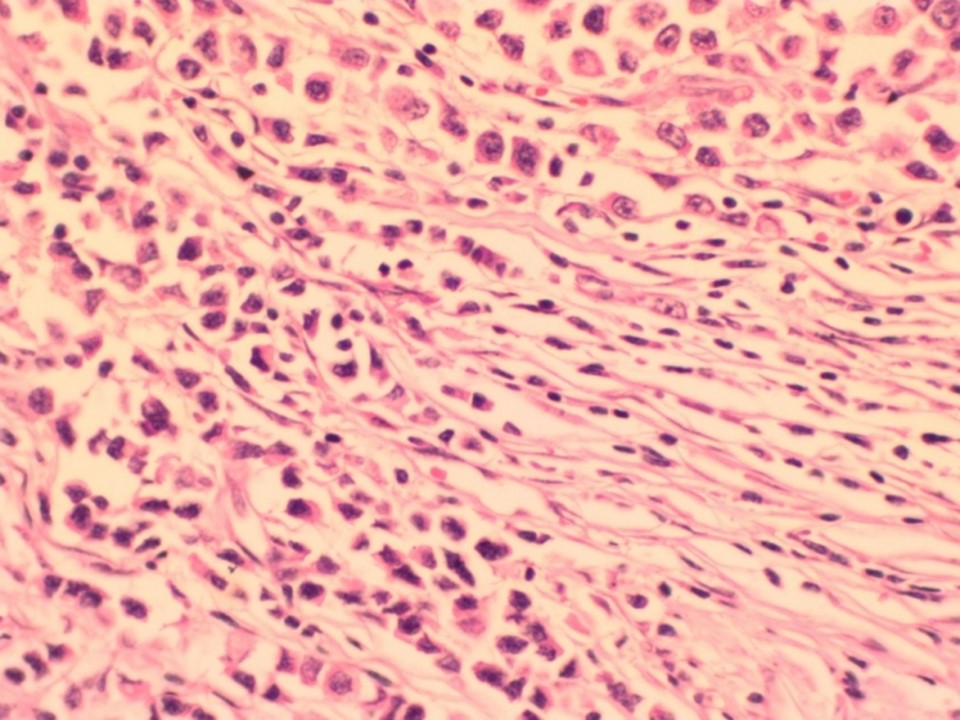 |
| Histopathology features: | |
| ‣ Specimen type: | MRM |
| ‣ Laterality: | Right |
| ‣ Macroscopy: | Cut surface shows a solid, whitish, firm to hard tumour (4.0 × 3.0 × 1.5 cm) with infiltrating margins. Tumour is situated at a depth of 2.0 cm below the skin. The base is 0.5 cm away from the tumour. A separate specimen labelled muscle from base was a single nodular brownish firm tissue bit (2.0 × 1.5 × 1.0 cm) |
| ‣ Histological type: | Infiltrating lobular carcinoma right breast |
| ‣ Histological grade: | Grade 1 (3 + 1 + 1 = 5) |
| ‣ Mitosis: | 4 |
| ‣ Maximum invasive tumour size: | 4.0 cm in greatest dimension |
| ‣ Lymph node status: | 5/30 |
| ‣ Peritumoural lymphovascular invasion: | Present |
| ‣ DCIS/EIC: | Absent; lobular carcinoma in situ not seen |
| ‣ Margins: | Free of tumour. Sections from the muscle from base show only skeletal muscle fibres and are free of tumour invasion |
| ‣ Pathological stage: | pT2N2 |
| ‣ Biomarkers: | |
| ‣ Comments: |
Case summary:
| Postmenopausal woman presented with right breast lump and right nipple retraction. Diagnosed as right breast carcinoma with skin and nipple retraction, BI-RADS 5 on imaging, as breast carcinoma on cytology, and as invasive lobular carcinoma, pT2N2 on histopathology. |
Learning points:
|